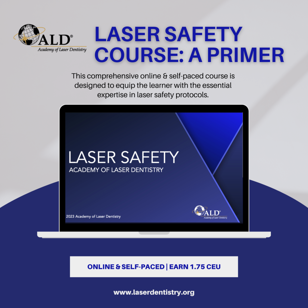
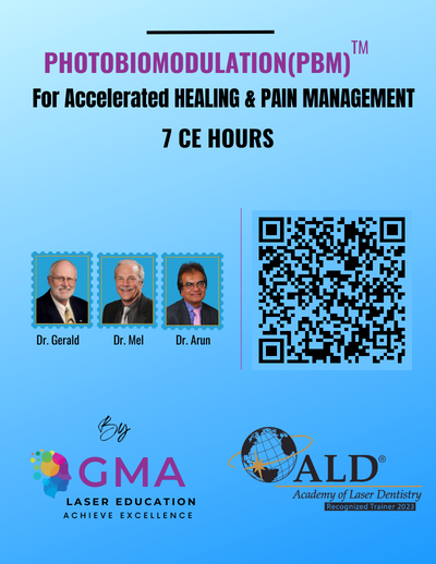
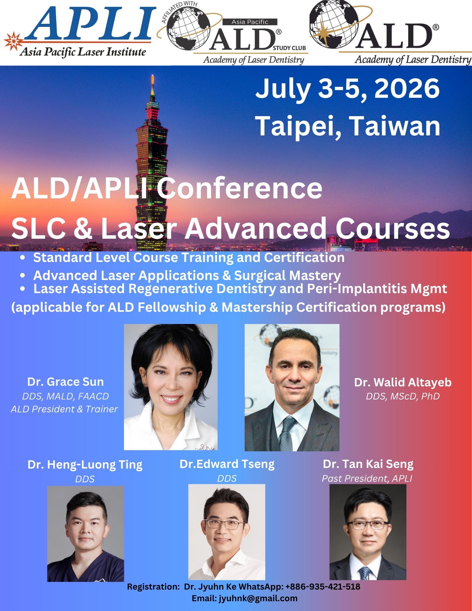
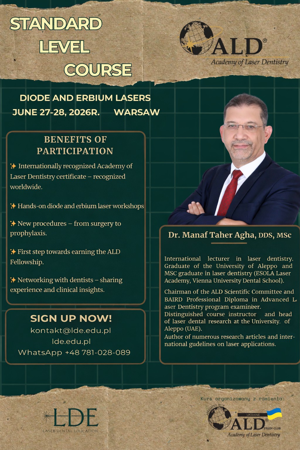
ALD 2026 COURSES
More to be announced
Advances in Laser-Assisted Regenerative Periodontal and Peri-Implant Plastic Surgery
Keynote Speaker: Homa H. Zadeh, DDS, PhDAdvances in laser technology have expanded the therapeutic options available for regenerative periodontal and peri-implant plastic surgical procedures. Lasers provide precise soft tissue ablation, improved hemostasis, reduced postoperative discomfort, and enhanced visualization of the surgical field, making them valuable adjuncts in procedures aimed at improving soft tissue phenotype and peri-implant tissue health. This presentation will review the application of laser technology in regenerative periodontal and peri-implant plastic surgery, with emphasis on soft tissue augmentation procedures. Clinical protocols for laser-assisted preparation of donor and recipient sites during connective tissue grafting and soft tissue phenotype modification will be discussed. The use of lasers to facilitate minimally invasive tissue management and optimize surgical outcomes will be highlighted. Non-ablative applications, including laser-assisted implant surface decontamination and photodynamic therapy for the management of periodontitis and peri-implantitis, will be reviewed. Clinical cases will illustrate the integration of laser technology into contemporary regenerative and plastic periodontal surgery, with discussion of the biologic rationale, surgical protocols, and practical considerations for optimizing treatment outcomes.
Learning Objectives:
- Describe the biologic principles and clinical indications for the use of laser technology in regenerative periodontal and peri-implant plastic surgery.
- Discuss the role of lasers in the preparation of donor and recipient sites during soft tissue augmentation procedures and in the management of surgical complications.
- Explain the application of non-ablative laser therapy, including implant surface decontamination and photodynamic therapy, in the treatment of periodontitis and peri-implantitis
Innovative Flapless Periodontal Surgery Using Er:YAG Laser: Er-LCPT
Keynote Speaker: Akira Aoki, DDS, PhDThe use of lasers in periodontal therapy has gained increasing attention because of their beneficial therapeutic and biological effects in the management of inflammatory and infectious periodontal diseases. Among the various laser systems available, the Er:YAG laser is particularly attractive due to its ability to safely and effectively treat both soft and hard tissues with minimal thermal damage.
In periodontal surgery, the Er:YAG laser can precisely debride diseased root surfaces, remove subgingival calculus, ablate inflamed soft tissues within periodontal defects, and promote favorable wound healing and periodontal regeneration. Clinical studies have demonstrated that Er:YAG laser-assisted periodontal surgery provides outcomes comparable to, and in some cases superior to, those achieved with conventional surgical approaches. However, despite numerous studies investigating laser-assisted periodontal pocket treatment, consensus regarding its clinical benefits has not yet been fully established.
This presentation introduces a novel minimally invasive flapless surgical approach, Er:YAG laser-assisted comprehensive periodontal pocket therapy (Er-LCPT), which combines Er laser irradiation with conventional mechanical instrumentation. Er-LCPT enables safe, effective, and comprehensive debridement of periodontal pockets without flap elevation. Clinical evidence, including case series and randomized controlled trials, has demonstrated the safety and efficacy of this procedure for the treatment of residual periodontal pockets, with significantly greater improvements in clinical parameters compared with scaling and root planing alone. Furthermore, Er-LCPT may serve as a flapless regenerative approach during initial periodontal therapy, offering new possibilities for minimally invasive periodontal treatment.
Learning Objectives:
- Describe the biologic principles and clinical indications for the use of laser technology in regenerative periodontal and peri-implant plastic surgery.
- Discuss the role of lasers in the preparation of donor and recipient sites during soft tissue augmentation procedures and in the management of surgical complications.
- Explain the application of non-ablative laser therapy, including implant surface decontamination and photodynamic therapy, in the treatment of periodontitis and peri-implantitis
Mastering the Parameters of PBM
Keynote Speaker: Jan Tunér, DDSPBM is a complex method including a great variation of available parameters. Looking casually at the litterature, it may seem that anything goes, but actually success always lies in strict observation of the parameters. In order to reach a professional consensus, studies need to be reproducable, but with inadequate or incorrect accounts of the used parameters, consensus is hard to reach. As a reviewer, it is often disappointing to see all the enthusiasm and time wasted by not being in control of the parameters before the very onset of a study. Our associations and journals need to offer potential researchers straighforward requirements in order to avoid such waste of efforts. The goal of this presention is to suggest the minimum requirements for a sound scientific article in order not to land in an endless revewing process or even rejection.
every procedure is a laser procedure!
Robin Horton, BDS, FALD, FWCLIThis talk is to show that lasers are not just for geeks doing specialised treatments. They are for everyday dentistry. The properties of dental lasers are such that they fit really well with almost everything we do. Lasers are your best friend in the operatory. We are a seven operatory Practice, and we have four erbium, dual tissue lasers and five diode, soft tissue lasers, because I want all my clinicians to be able to use a laser every day in their dentistry. During the presentation I will give an overview and insight into laser and digital dentistry, how laser fits really well with the digital dentistry of today. I will show what the digital workflow is and how it streamlines procedures and simplifies solutions. I will show what is out there in terms of digital devices and what they bring to the table. We will run the whole range of dental procedures finishing with the current pinnacle in implant dentistry of how it is now possible to scan, plan, extract, place, get accurate records, print and fit teeth, all in a few hours and what role laser has in this which makes the procedure so much more predictable. But aside from this highly technical procedure Lasers are amazing in fillings, perio, soft tissue surgery, extractions, bone removal, sectioning roots, removing infection, raising flaps, uncovering implants, endodontics, healing, reducing inflammation, I could go on!
Learning Objectives:
- To understand the full range of dental treatments possible with a dental laser
- To understand what the digital workflow is and how lasers fit in with this way of working
- To see how we use a dual tissue laser instead of conventional tools to minimise damage and pain to the patient and increase patient satisfaction
- To see just how predictable immediate implant placement following laser extraction can be, using guided, minimally invasive, laser assisted surgery
Innovative approaches in managing oral biofilm and periopathogens using laser therapy
Kinga Grzech-Leśniak, Prof., DMD, MSc, PhDThe management of oral biofilm and periopathogens is a critical aspect of periodontal therapy, influencing treatment outcomes and long-term oral health. Traditional mechanical debridement, while effective, has limitations in eradicating pathogenic bacteria, particularly in deep periodontal pockets and complex biofilm structures. Laser therapy presents an innovative approach by offering bactericidal effects, biofilm disruption, and biostimulatory benefits that promote tissue healing. This presentation explores the role of various laser systems, including Nd:YAG, Er:YAG, and diode lasers, in modulating the oral microbiome and enhancing periodontal regeneration. Clinical applications, treatment protocols, and the latest evidence-based findings on laser-assisted periodontal therapy will be discussed, emphasizing its advantages over conventional methods.
Learning Objectives:
- Understand the role of laser therapy in biofilm management
- Evaluate the clinical applications of laser-assisted periodontal therapy
- Assess the benefits of laser therapy beyond bacterial reduction
laser to legacy: converting clinical excellence into long-term wealth
Nick Clausen, MBAMany laser dentists create best-in-class clinical outcomes and patient experiences—yet the practice production does not always reflect that level of excellence. When production, collections, and EBITDA depend entirely on the systems and teams the owner builds, poor structure silently suppresses valuation and eliminates freedom. This presentation is specifically designed for the owner-doctor who wants to transition from primary producer to Dental CEO and ultimately build an enterprise that delivers maximum value and time freedom. This is not something that can be engineered one year before retirement—the foundation must be built early and intentionally. Attendees will learn how to shift from working in the practice to working on the business by adopting a CEO mindset centered on communication, leadership, accountability, and scalable profitability. We will explore growth strategies that expand revenue without adding clinical days, including the four revenue streams of a successful dental enterprise. Participants will gain clarity around the Key Performance Indicators (KPIs) that influence valuation, how to structure accountable leadership within the team, and the critical differences between being a boss and becoming an effective leader. Practical systems will be provided for conflict resolution, corrective communication, and internal leadership development. The goal is simple: build a practice that thrives without you at the center—so when you are ready to exit, your business commands premium value and creates long-term wealth.
Learning Objectives:
- Implement communication structures that prevent production leakage and operational drift
- Implement CEO leadership and communication frameworks that build accountable, self-managing teams
- Recognize and track the critical KPIs that directly impact profitability, enterprise value, and exit readiness
- Understand that four ways how CEO's that increase scalability, practice valuation, and EBITA are traditionally compensated
When Evidence Falls Short: Rethinking How We Interpret Research in Laser Dentistry
Ali Obeidi, DDS, MSc, MS, FALDEvidence-based dentistry has become an essential foundation of modern clinical decision-making. However, confusion often arises when clinicians encounter procedures that appear to work reliably in practice but lack extensive randomized clinical trials. This situation is particularly common in laser dentistry, where technological innovation frequently progresses faster than the research needed to formally document every clinical application. This presentation introduces a well-known satirical example from medical literature in which researchers humorously demonstrated that no randomized trials existed proving that a life-saving device actually prevents death when jumping from an aircraft. The example illustrates an important lesson in scientific interpretation: the absence of published evidence does not necessarily mean that a technique is ineffective. This concept is highly relevant to laser dentistry. Many laser-assisted procedures—such as soft-tissue surgery, bacterial reduction, implant surface decontamination, and photobiomodulation for pain control—are widely used by clinicians who consistently observe positive outcomes. Yet these applications are sometimes dismissed simply because large-scale trials or standardized protocols are still evolving. Using this analogy as a starting point, this presentation encourages clinicians to look at scientific literature with a broader perspective—recognizing both the value and the limitations of published evidence while considering biological plausibility and real-world clinical outcomes. The goal is to stimulate thoughtful discussion about how innovation, clinical experience, and research can work together to strengthen the scientific foundation of laser dentistry.
Learning Objectives:
- Recognize the difference between absence of evidence and evidence of ineffectiveness
- Apply critical thinking when interpretting scientific literature related to laser dentistry
- Understand how clinical experience, biological plausibility, and research complement evidence-based practice
Use of Erbium Lasers for Debonding
Janina Golob Deeb, DMD, MSRemoval of all-ceramic restorations can be time consuming and challenging. Erbium lasers offer an alternative noninvasive method for removal of ceramic restorations from implants and teeth, as well as fiber posts and brackets. Erbium lasers used for debonding include Er:YAG and Er,Cr;YSGG. They are both effective noninvasive tools and may be considered as a viable alternative to using rotary instrumentation for removal of ceramic restorations without causing harm to abutments or prosthesis. The retrieval process by laser does not destroy the restorations, thus they can be used as temporaries or recemented in improved position following laser-assisted debonding. This lecture aims to summarize the research findings and clinical examples using erbium lasers for debonding of various restorations and appliances from natural teeth and dental implants. Effective protocols for various ceramics and cement types will be discussed supported by current evidence and laser parameters. Clinical cases include debonding of veneers, single crowns and fixed partial dentures of natural teeth and implant abutments cemented with various cement materials. Laser settings affect the debonding time and the increase in temperature. Examination under magnification of debonded ceramics demonstrates minimal structural effects resulting from laser applications when appropriate parameters are used. When used incorrectly, side effects may include temperature rise and damage to tooth structures. Duration of procedure to retrieve a ceramic restoration is influenced by type of ceramic, its translucency and thickness, surface area of abutment and restoration, type of restoration and its retentive features, and types and thickness of cement used.
Learning Objectives:
- Understand which wavelengths are best suited for debonding of ceramic restorations
- Discuss evidence-based protocols for laser-assisted ceramic debonding with Erbium lasers
- Understand the properties of restorations and cements that influence debonding efficiency
- Select the appropriate laser parameters for debonding ceramic restorations
Laser-Assisted Implantology: Improving Precision, Stability, and Healing in Implant Treatment
Saleh Anvaria Aria, DDS, MScDental implant therapy has become a predictable treatment modality; however, achieving optimal precision during surgery, maximizing primary stability, and promoting rapid soft- and hard-tissue healing remain key determinants of long-term success. The integration of dental laser technology into implantology has introduced new opportunities to enhance surgical accuracy, minimize tissue trauma, and improve postoperative outcomes. This lecture will explore the clinical role of laser-assisted implantology across multiple stages of implant treatment, including soft-tissue management, bone preparation, implant site decontamination, and peri-implant tissue modulation. Particular emphasis will be placed on the use of all-tissue laser systems, such as the Er,Cr:YSGG 2780 nm wavelength, which enables precise interaction with both soft and hard tissues while preserving surrounding biological structures. The presentation will discuss how laser energy can facilitate minimally invasive surgical approaches, enhance visibility through improved haemostasis, reduce bacterial contamination, and stimulate photobiomodulation effects that support faster healing and improved patient comfort. Clinical cases will demonstrate the application of lasers in immediate implant placement, implant site preparation, peri-implantitis management, and soft-tissue contouring. Evidence-based principles, laser-tissue interaction mechanisms, and practical clinical protocols will be reviewed to help clinicians integrate laser technology safely and effectively into implant treatment workflows. By combining advanced laser technology with modern implant concepts, clinicians can achieve greater surgical precision, improved implant stability, and enhanced biological healing responses.
Learning Objectives:
- Understand the principles of laser-tissue interaction relevant to implant surgery
- Identify the clinical advantages of laser-assisted implantology in both soft- and hard-tissue procedures
- Apply laser protocols for implant site preparation, soft-tissue management, and peri-implant decontamination
using Lasers to Help Diagnose and Manage Auto-Immune Diseases
Laura Braswell, DDSAuto-immune diseases are becoming more common since the advent of Covid. Often called silent diseases, the patients often do not appear ill in general, but suffer quietly with pain and lifestyle limitations. Lasers can be helpful for both ablative treatments and photobiomodulation for healing and pain control. This presentation will help the practitioner with diagnostic skills to realize the signs and symptoms of these diseases as well as the multiple possible etiologies. The use of lasers can be life-changing for patients and eleviate much anxiety and suffering.
Learning Objectives:
- Formulate a differential diagnosis for auto-immune diseases and determine what tests are appropriate to assist in confirmation
- To determine what lasers are appropriate for treatment and palliative care for these patients
- Case presentations will be provided to demonstrate each objective
Laser Dentistry as a Transformative Educational Tool: Opportunities and Barriers in Academic Integration
Natalia Elson, DDSThe rapid evolution of dental technology requires academic leadership to ensure that graduates are prepared to deliver minimally invasive, technology-driven care. Laser dentistry represents both an advanced clinical modality and a strategic educational tool that can enhance student engagement, clinical competence, and interdisciplinary learning. This presentation evaluates the integration of laser dentistry into dental curricula through a structured, outcomes-based educational framework. A phased implementation model is proposed, beginning with foundational didactic instruction focused on laser physics, safety, and tissue interaction, followed by simulation-based training to develop psychomotor skills, and culminating in supervised clinical application. Integration within existing disciplines—including periodontics, restorative dentistry, and oral surgery—allows efficient incorporation without increasing curricular burden. Defined competencies, standardized safety certification, and objective assessment methods support consistency and measurable student outcomes. Educational leadership plays a critical role in successful implementation. Faculty development, calibration, and mentorship ensure alignment across departments and promote confidence in teaching laser applications. Administrative support and resource allocation further enable sustainable integration. Measurable outcomes include increased student participation in laser-assisted procedures, improved clinical decision-making, enhanced patient communication, and greater readiness for contemporary practice. Key challenges include financial investment, curriculum time constraints, and resistance to change; however, these can be addressed through phased adoption, interdisciplinary collaboration, and structured faculty training programs. Laser dentistry serves as a catalyst for modernizing dental education. Through intentional curriculum design and leadership-driven implementation, academic institutions can produce graduates who demonstrate competency in emerging technologies, apply evidence-based clinical reasoning, and deliver patient-centered care aligned with current standards of practice.
Learning Objectives:
- Define core competencies required for integrating laser dentistry into undergraduate and graduate dental curricula.
- Describe a phased, outcomes-based model for implementing laser education, including didactic, simulation, and clinical training componenets.
- Analyze common barriers to curriculum integration and select evidence-based strategies for faculty development, resource allocation, and program organization.
- Assess the impact of laser education on student performance, including clinical decision-making, procedural confidence, and patient-centered care outcomes.
Utilizing Photobiomodulation (PBM) to Treat Cancer Symptoms and Cancer Treatment Side Effects
Mel A. Burchman, DDS, MALDAs we all know cancer is a devastatingly painful desease, both in it's symptoms and unfortunately also during it's treatment. Some of the cancer treatent side effects are so painful that patients just want to stop their ttreatments and die in peace. Over many years of practice I have treated many patients, friends and family members that are experiencing this horrible desease, from a young girl of 3 to senior citizens. During this time I have developed several techniques for treating the symptoms and side effects of this dreaded desease. I also have had the joy of helping these people in their time of great pain. As a matter of fact, at this time I am helping a dear friend who is battling breast cancer. I have taught her how to treat her pain and symptoms and have leant her one of my lasers and it just makes me feel wonderful when I speak to her and she tells me she is using the laser everyday and it really is helping her. Several years ago I was sent a patient by Dr. Chip Whitney that had colon and liver cancer and said the first round of chemo and radition were so painful he wanted to discontinue treatment. Dr. Whitney talked him in to seeing me saying, "what do you have to lose.' Well the treatment results were excellent and the patient completed his treatment. Dr. Whitney. wrote an article about me and my treatment in the Academy of Oral and Systemic Health Journal and said, "My patient said the results of the laser treatment were LIFE CHANGING" and "Now thanks to my recommendation my patient will complete his treatment" On a more personal note, I was able to treat and help my wife Sue during her 3 year bout with advanced pancreatic cancer. I love helping these people and want to teach others how to preform these treatments for nausea, digestive problems, neuropathy, cold fingers, loss of taste, headaches and oral mucositis
Learning Objectives:
- During this course the attendee will learn how to use PBM to treat neuropathy
- The attendee will learn how to treat a cancer patient suffering from nausea and digestive problems
- Many cancer patients suffer from headaches and TMJ pain, by attending this presentation the student will learn how to treat these problems
- Oral mucositis is considered one of the most debilitating side effects of cancer treatment, the attendee will learn how to treat OM with photobiomodulation (PBM) techniques
From Perforation to Preservation: A Laser-Led Amazing Human Body Regeneration, Laser-Assisted Multimodal Management of an Extensive Mandibular Periapical Granuloma with Cortical Plate Perforation and Risk of Pathological Fracture
Mohamed Yehya, DDS, PhDBackground: Extensive periapical lesions with cortical plate perforation and advanced mobility are often considered hopeless and treated by extraction, particularly when structural compromise raises concern for pathological fracture. Laser-assisted protocols may enhance disinfection, surgical precision, and postoperative healing, potentially allowing preservation of compromised teeth.
Case Presentation: A medically fit patient presented with persistent purulent discharge from an extraoral chin sinus tract, chronic discomfort, and mobility of mandibular anterior teeth. Previous consultation recommended extraction of teeth 32–43 due to severe bone destruction and risk of pathological fracture. Clinical examination revealed Grade III mobility in tooth 31, Grade II mobility in 32, 41, and 42, and Grade I mobility in 43. CBCT demonstrated a large, well-defined radiolucent lesion measuring 21.5 mm mesiodistally and 13.7 mm superoinferiorly, extending from teeth 32 to 43. The lesion perforated the labial cortical plate and extended lingually, leaving less than 1 mm thickness of the lingual cortex. All involved teeth were non-vital.
Intervention: Initial therapy included scaling and root planing, emergency access, chemomechanical preparation, intracanal medicament placement, and laser-assisted disinfection. Photodynamic therapy using a green photosensitizer activated with an 810-nm diode laser (300-µm fiber) was applied intracanal and to periodontal pockets. The teeth were splinted prior to surgical intervention to enhance stability. Surgical management involved full-thickness flap reflection, extensive curettage, apicectomy, and retrograde mineral trioxide aggregate filling. Adjunctive 810-nm photothermal irradiation was performed to eliminate residual granulation tissue prior to grafting. The defect was reconstructed using platelet-rich fibrin combined with particulate bone allograft, followed by postoperative photobiomodulation therapy.
Results: Histopathological analysis revealed chronic inflammatory granulation tissue consistent with a periapical granuloma, confirming the endodontic origin of the lesion. Early follow-up demonstrated sinus resolution, improved stability, and favorable soft tissue healing. Radiographic monitoring is ongoing to evaluate bone regeneration.
Conclusion: A laser-integrated, multidisciplinary approach enabled preservation of teeth previously deemed hopeless despite extensive cortical perforation, highlighting the potential of advanced laser protocols in managing large inflammatory periapical lesions.
Learning Objectives:
- Role of diode laser in enhancing disinfection, surgical precision, and postoperative healing, potentially allowing preservation of compromised teeth.
- Different concepts of laser assisted interventions and approaches Photobiomodulation, Photothermal, Photodynamic therapies.
- Secrets behind different laser powers and dual action of both powers high to kill and lower to regenerate, cumulative effects and precise dosimetry.
- Combination of low power and PRF as innovated approach for regeneration, (Photosynthesis) every thing grows by light.
Thermal Imaging–Monitored At-Home Photobiomodulation Therapy for Orofacial Inflammatory Processes
Haitham Elafifi, DDSBackground: Orofacial inflammatory conditions, including temporomandibular disorders, post-surgical inflammation, neuropathic pain, and mucosal lesions, are frequent clinical challenges that significantly affect patient comfort and quality of life. Photobiomodulation therapy (PBMT) has gained increasing attention as a non-invasive treatment modality capable of modulating inflammation, reducing pain, and promoting tissue healing. Recent advances in portable light-based devices have enabled the application of PBMT in home settings, potentially expanding treatment accessibility and patient autonomy.
Objective: This presentation explores the potential integration of at-home photobiomodulation therapy with infrared thermal imaging as a monitoring tool for orofacial inflammatory processes.
Concept and Methods: Infrared thermography allows the detection of subtle variations in skin temperature associated with vascular and inflammatory changes. Sequential thermal imaging of the orofacial region may provide a non-invasive method to monitor inflammatory activity, identify thermal asymmetries, and evaluate response to PBMT over time. The combination of home-based PBMT protocols with periodic thermal imaging could allow clinicians to remotely monitor treatment progress and adjust therapeutic parameters when necessary.
Clinical Relevance: Integrating photobiomodulation therapy with thermal imaging monitoring may provide a personalized and technology-assisted approach for the management of orofacial inflammation, potentially reducing the need for frequent clinical visits while maintaining objective follow-up.
Conclusion: At-home PBMT combined with thermal imaging monitoring represents a promising strategy for the management of orofacial inflammatory conditions. Further clinical research is needed to establish standardized protocols and validate its clinical effectiveness.
Learning Objectives:
- Understand the principles and clinical mechanisms of photobiomodulation therapy (PBMT) in the management of orofacial inflammatory conditions, including its effects on inflammation modulation, pain reduction, and tissue healing.
- Recognize the role of infrared thermal imaging as a non-invasive diagnostic and monitoring tool for detecting thermal asymmetries and inflammatory activity in the orofacial pain.
- Evaluate the potential integration of at-home photobiomodulation therapy with thermal imaging monitoring as a strategy to support personalized treatment protocols and remote follow-up in patients with orofacial inflammatory processes.
Effect of Photobiomodulation Therapy on Postoperative Pain and Trismus Following Implant-Related Oral Surgeries: A Systematic Review of Randomized Controlled Trials
Nahid Derikvand, MDS, Laser FellowshipBackground: Postoperative pain and trismus remain common complications following implant-related oral surgeries, potentially affecting patient comfort and recovery. Photobiomodulation therapy (PBM) has been proposed as a non-invasive adjunctive approach to modulate inflammation and enhance postoperative healing; however, its clinical effectiveness in implant-related procedures remains controversial.
Objective: This systematic review aimed to evaluate the effect of photobiomodulation therapy on postoperative pain and trismus following implant-related oral surgeries based on evidence from randomized controlled trials.
Methods: A systematic literature search was conducted in PubMed and ScienceDirect for randomized controlled trials assessing the effects of PBM after implant-related oral surgical procedures. Studies evaluating postoperative pain, trismus, or mouth opening were included. Data extraction and qualitative synthesis were performed in accordance with PRISMA guidelines.
Results: Nine randomized controlled trials met the inclusion criteria. Most studies reported a significant reduction in postoperative pain intensity in PBM-treated groups compared with controls. Limited but consistent evidence suggested a beneficial effect of PBM on trismus and maximal mouth opening, particularly in the early postoperative period. Considerable heterogeneity was observed in laser parameters, treatment protocols, and outcome assessment methods.
Conclusion: Current evidence suggests that photobiomodulation therapy may reduce postoperative pain and, to a lesser extent, trismus following implant-related oral surgeries. However, variability in treatment protocols and methodological limitations among existing trials highlight the need for well-designed, standardized randomized controlled studies to establish optimal PBM parameters and confirm its clinical effectiveness.
Keywords: Photobiomodulation therapy; Low-level laser therapy; Dental implants; Postoperative pain; Trismus; Oral surgery
Learning Objectives:
- Evaluate the effect of photobiomodulation therapy on postoperative pain after oral surgery
- Evaluate the effects of photobiomodulation therapy on trismus following oral surgery
- Evaluate the effects of photobiomodulation therapy on trismus and pain following advanced implant surgery
Laser-Assisted Management of Severely Curved Canals to Prevent Ledge Formation
Md Arifur Rahman, BDS, MScIntroduction: The chemo-mechanical preparation of root canals with severe curvatures presents a significant clinical challenge, often predisposing the tooth to iatrogenic procedural errors such as ledge formation, canal transportation, and apical zipping. A ledge prevents adequate disinfection and obturation of the apical third, which is linked to persistent periapical infection. Recently, diode laser-assisted endodontics has emerged as a promising adjunct to traditional mechanical preparation.
Case Presentation: A clinical case involving a mandibular molar with a severely curved mesial root is presented. To avoid ledge formation and preserve the original canal anatomy, mechanical instrumentation was minimized, and diode laser-activated irrigation was utilized to facilitate deep dentinal disinfection and smear layer removal. Radiographic evaluation demonstrated successful negotiation, cleaning, and a dense three-dimensional obturation seamlessly following the natural canal curvature.
Conclusion: The integration of diode laser technology in the management of complex canal curvatures safely enhances irrigant penetration and disinfection, reduces the necessity for aggressive mechanical filing, prevents ledge formation, and ensures a highly adapted root canal seal.
Combination Laser Therapy—Effectively Using Multiple Wavelengths for Best Overall Treatment Results
Maki Tanaka, DDS, PhDAdvantages of Laser treatments are:
- Decreasing the risk of contamination by sterilization /disinfection of bacteria.
- Minimizing damage to healthy surrounding tissue.
- Less discomfort/pain pre-op and post-op.
- Less bleeding, reduced inflammation and faster/better healing.
With 20 years of experience with dental lasers in my practice, the use of lasers makes difficult cases easier with the ability to precisely and accurately perform treatments resulting in merits for both patients and doctors. In addition, selectively combining multiple laser modalities with different wavelengths allows for customized treatments and increased accuracy. For periodontal treatment, 635nm diode laser can be used for a-PDT to disinfect the periodontal pocket or surgical sight post op treatment. Combined with 2940nm Er:YAG laser for areas biologically difficult to access such as deep pocket or root furcation in non-surgical procedures or debridement in surgical procedures. NIR lasers can be used for PBM to induce a faster, better healing process. I will be discussing the importance of understanding characteristics of each laser wavelength and share some of my cases combining multiple wavelengths for overall customized treatment.
Learning Objectives:
- What wavelength of laser and how should it be used to most effectively eliminate the source of infection?
- To accelerate wound healing, which wavelength of laser and how should it be used most effectively?
- What are the clinical benefits of combined laser therapy?
Laser-Induced Hypoxia of the Post-Extraction Socket to Preserve Bone Volume Without Grafting
Ernesto Vatteroni, DDS, PhDObective: Post-extraction alveolar sockets undergo significant dimensional resorption, losing up to 30% of bone volume within one year. This is particularly critical in the anterior maxilla, where delayed implant placement and prosthetic rehabilitation lead to compromised esthetic outcomes. Previous studies have demonstrated that immediate implant placement with provisionalization reduces socket contraction, a phenomenon linked to transient hypoxia: sealing the socket limits oxygen diffusion, triggering a biochemical cascade that promotes neoangiogenesis, osteoblast activation, and early inhibition of myofibroblasts — resulting in reduced bone resorption and preserved soft tissue volume. Building on this mechanism, this study investigates whether diode laser application alone can induce a comparable state of transient hypoxia in post-extraction sockets, preserving ridge dimensions without any bone grafting or implant placement.
Materials and Methods: Three non-smoking, systemically healthy patients (2 males, 1 female; ages 33, 54, and 61) who declined immediate implant placement and provisional restorations were enrolled. All extractions involved the maxillary first premolar (esthetic zone). Pre-operative records included CBCT (DICOM files) and intraoral digital scans (STL files). Following atraumatic extraction, a diode laser (980 nm, non-initiated tip, non-contact mode, hemostasis setting) was applied over the blood clot for 2 minutes, with the aim of creating a superficial vitrified layer to seal the socket and induce transient hypoxia. No grafting materials or membranes were used. At 4 months post-extraction, new CBCT and intraoral scans were acquired. Dimensional changes were assessed by superimposition of pre- and post-operative STL files (soft tissue and ridge contour analysis) and DICOM files (bone volume and hard tissue analysis), allowing quantification of osseous, soft tissue, and total alveolar volumetric changes. At the time of implant placement, a core biopsy was obtained for histological evaluation of bone quality and quantity.
Results: All three cases showed consistent outcomes. Volumetric bone contraction averaged 5%, while soft tissue contraction averaged 3% — both substantially below the values reported in the literature for ungrafted, unsealed sockets. At the time of implant placement, the socket was completely filled with newly formed bone. Histological analysis revealed partially trabeculated bone with adequate quality for implant osseointegration.
Discussion and Conclusion: Although the sample size is limited (n=3), the consistency of results across all cases is encouraging and supports the hypothesis that diode laser-induced superficial vitrification of the blood clot generates transient hypoxia, thereby inhibiting myofibroblast contraction and stimulating osteogenic activity. This represents a simple, minimally invasive technique — a single 2-minute laser application — requiring no grafting materials. These preliminary findings warrant further investigation through larger controlled clinical trials. Laser-induced hypoxia may represent a paradigm shift in post-extraction socket management, offering a straightforward and accessible protocol for every trained laser clinician.
Learning Objectives:
- Explain the biological mechanism of laser-induced transient hypoxia in post-extraction sockets and its role in inhibiting myofibroblast contraction and promoting osteogenesis.
- Describe the clinical protocol for diode laser application (980 nm, non-initiated tip, non-contact, hemostasis mode) following tooth extraction to seal the socket and preserve alveolar bone volume without grafting.
- Evaluate the use of STL and DICOM file superimposition as a method to quantify hard and soft tissue dimensional changes following post-extraction laser treatment.
- Assess the potential of laser-induced hypoxia as a miniamlly invasive alternative to bone grafting for ridge preservation in the esthetic zone, based on preliminary histological and volumetric evidence.
Photobiomodulation Therapy Using 890 nm Diode Laser in Management of Temporomandibular Joint Disorder—A Prospective Longitudinal Clinical Study
Ujjwal Prem, MDSObjective: Efficacy of 980 nm diode laser in photo biomodulation therapy for management of temporomandibular joint disorder.
Purpose: Temporomandibular joint disorder (TMD) encompasses a spectrum of clinical signs and symptoms affecting the masticatory muscles, the temporomandibular joint (TMJ), and associated structures. A wide range of therapeutic approaches have been proposed to alleviate or resolve patient symptoms, particularly pain, jaw deviation, mouth opening and joint sounds such as clicking. Photobiomodulation/Low-level laser therapy (LLLT) has been utilized for analgesia and to promote tissue healing, although its efficacy in TMD management remains controversial due to limitations in existing evidence. Therefore, the present study aims to evaluate the effectiveness of 980 nm LLLT in patients with TMD.
Materials and Methods: A prospective longitudinal clinical trial was conducted on 150 patients diagnosed with temporomandibular joint disorder (TMD). Baseline matching was performed for gender, pain intensity, mouth opening, jaw deviation and joint clicking on day1.Low-Level Laser Therapy (LLLT) was administered using a 980 nm wavelength at 10kHz, delivering a total energy of 120 J over three TMJ points (40J per point), along with 120 J applied to additional sites of muscle tenderness, for a duration of 1 minute.Software: Microsoft Excel and Jamovi (v2.6).Descriptive: Continuous variables (Mean ± SD); Categorical variables (Frequency/Percentage).Categorical (Jaw sounds/deviation): Cochran’s Q test was used across four time points (Days 1, 3, 7, 14) followed by McNemar’s test with Bonferroni correction for pairwise comparisons.Continuous (Mouth opening): Repeated Measures ANOVA was used for longitudinal changes, with Tukey’s test for post hoc comparisons.Inter-group (VAS scores): Independent samples t-test compared patients with vs. without dental prostheses at each time point.A p-value of< 0.05 was considered statistically significant.
Results: Changes in jaw sounds at baseline (Day 1), 68 patients (45.3%) exhibited jaw sounds, which progressively decreased to (15.3%) on Day 7, and completely resolved by Day 14.Jaw deviation at baseline (Day 1), 50 patients (33.3%) exhibited jaw deviation, which decreased to (4.7%) on Day 7, with complete resolution observed by Day 14 where all patients (100%) had no jaw deviation. Analysis using Cochran's Q test and McNemar's test with Bonferroni correction.The mean mouth opening increased progressively from 41.29 ± 7.10 mm on Day 1 to 46.43 ± 2.22 mm on Day 14. Analysis using Repeated Measures ANOVA revealed a statistically significant difference in mean mouth opening across the time intervals (p< 0.001).Post hoc analysis using Tukey’s test Overall,these findings indicate a progressive and statistically significant improvement in mouth opening following laser therapy,with the greatest increase observed by Day 14. The comparison of VAS scores based on the presence of dental prostheses at baseline, the mean VAS scores were slightly lower in patients without prostheses (5.04 ± 1.14) compared to those with prostheses (5.37 ± 1.04).
Conclusion: Laser therapy is an effective intervention for the total resolution of jaw sounds and deviation. It significantly improves functional mouth opening and provides substantial pain relief. While the presence of dental prostheses may slightly modulate the rate of pain reduction. Keywords :- LLLT, temporomandibular joint disorder, diode laser, jaw sounds.
Learning Objectives:
- Study helps in identifying the clinical parameters of Temporomandibular Joint Disorder (TMD)—specifically jaw sounds, deviation, and restricted mouth opening—in photobiomodulation.
- Analyzing the longitudinal efficacy of LLLT in achieving total resolution of joint clicking and jaw deviation.
- The impact of dental prostheses on the rate of pain reduction (VAS scores) when implementing laser therapy for TMD patients.
lasers for the GP and beyond
Larry Lieberman, DDSYou need to learn about lasers—and here's why. For more than three decades, I have incorporated laser technology into my daily dental practice, using it throughout the day for a wide range of procedures, many of which would be difficult or impossible to perform without it. Laser dentistry benefits not only patients through improved comfort and outcomes, but also enhances team engagement, practice efficiency, and profitability. Integrating laser technology can help elevate your practice to a true state-of-the-art level.
While laser-assisted tooth preparation is often what first comes to mind, it is only the beginning. Discover why lasers have become one of the most effective tools for treating periodontal disease, peri-implantitis, root canal infections, and many other clinical conditions. Learn about photobiomodulation and how this exciting technology can promote healing, reduce inflammation, and provide pain relief. Explore advanced laser applications in facial esthetics that can help reverse visible signs of aging, as well as innovative procedures such as NightLase®, which can reduce snoring and improve symptoms associated with sleep apnea.
With more than 40 years of clinical dental experience and over 30 years of laser expertise, I will share practical, real-world techniques that can be immediately incorporated into your practice to improve patient care and expand treatment possibilities.
Learning Objectives:
- I am looking forward to sharing my passion!
- Show how lasers can make your practice better and your patients happier
- I will present another revenue stream to increase your bottom line
Integrated Photonics-Enhanced Precision Dentistry—Current Status and Future Trends of Applications Across Dental Disciplines
Ambrose Chan, PhD, MSc, BDS, FRACDS, FICDPhotonics has superseded traditional electronics and is revolutionizing dentistry in the 21st century through its precision, speed, efficiency, versatility, and enhanced productivity. The growing emphasis on energy-efficient, streamlined, and patient-oriented applications aligns with strong market growth, with the global photonics market projected to more than double in value within the next decade to USD 1700 billion (Marketresearchfuture, 2025).
Dental photonics encompasses the application of photon-based energy sources (e.g. lasers, diode lasers, light-emitting diodes: LEDs and broadband light…etc.) and has become an increasingly integral component of modern clinical practice, contributing to improved patient experience with minimally invasive and regenerative-oriented dentistry while demonstrating measurable value in enhancing clinical efficiency and productivity. The integration of photonics and cone-beam computed tomography (CBCT) and digital dentistry (optical scanning, manufacturing, and radiography) has further transformed clinical practice by expanding diagnostic capabilities, streamlining workflow, enhancing procedural precision and predictability across dental disciplines. This review highlights and summarizes the merits and major technological advancements of the current most widely accepted and evidence-supported clinical applications of integrated photonic dentistry, and discusses emerging evidence and future trends across all dental disciplines. In conclusion, integrated dental photonics represents a rapidly evolving future central framework to converge digital-supported, artificial intelligence (AI)-enabled, robotic-guided and personalized theranostic technologies into an integrated precision oral healthcare ecosystem.
This presentation will give a highlight of the merits and major technological advancements of the current most widely accepted, evidence-supported clinical applications of integrated dental photonics, and future trends across dental disciplines.
Learning Objectives:
- Understanding integrated photonic dentistry
- Summarizing the merits and technological advancements of the current most widely accepted, evidence-supported clinical applications of integrated photonic dentistry
- Describing the future trends in dental photonic integration
Dental Hygienist Track Courses
More to be announced
"Lighting the Way" photobiomodulation for healing, pain, and health in hygiene
Lynn Atkinson, RDHPhotobiomodulation (PBM), formerly known as low-level laser therapy, is emerging as a powerful adjunct in periodontal and implant care. By delivering low-dose red and near-infrared light to tissues, PBM can modulate mitochondrial function, reduce inflammation and pain, and enhance wound healing without cutting or ablating tissue. This course translates the science into practical chairside protocols tailored to periodontists, hygienists, and restorative clinicians. We will review current evidence for PBM in periodontal wound healing, post-operative pain, oral mucositis, TMD/orofacial pain, and peri-implant therapy, along with real-world parameters for diode lasers commonly used in practice. Attendees will leave with a clear framework for integrating PBM into existing workflows—from diagnosis and case presentation to post-op care and maintenance.
Learning Objectives:
- Explain the core mechanisms of photobiomodulation at the cellular level (mitochondria, cytochrome c oxidase, signaling cascades)
- Learn how to calculate appropriate PBM parameters (wavelength, power, spot size, time, fluence) for common periodontal applications using diode lasers
- Communicate the value of PBM to patients and the interdisciplinary team using clear, evidence-based language
Deferring and Referring with Confidence”: Clinical Judgment, Ethical Responsibility, and Collaborative Care Decision-Making for the Dental Team
Erin Doffoney, RDHThe increasing integration of laser-assisted periodontal therapy into dental hygiene and general practice settings has expanded nonsurgical treatment capabilities while simultaneously increasing the need for evidence-based case selection and interdisciplinary referral decision- making. This presentation examines clinical indicators that distinguish patients who may benefit from conservative laser-assisted periodontal therapy from those who require comprehensive periodontal evaluation and specialty intervention. Emphasis will be placed on assessment of periodontal staging and grading, persistent inflammation, refractory disease patterns, furcation involvement, mobility, osseous defects, peri-implant complications, systemic risk factors, and patient-specific considerations that influence prognosis and treatment outcomes. The current literature on laser-assisted periodontal therapy, risk assessment protocols, and collaborative management strategies will be reviewed to support ethical and patient-centered clinical decisions. Attendees will gain practical guidance on recognizing the limitations of nonsurgical therapy, improving communication with periodontal specialists, and developing referral protocols to enhance long-term periodontal stability and patient care outcomes.
Are the Rules and Regulations for Lasers in Dental Hygiene Changing Again?
Angie Wallace, RDHIn this presentation, the attendees will see the States that allow Hygienists to use lasers, along with what the verbiage in each State lists. We will visit the hours needed for a hands on requirement, as well as the understanding of what it means to have training, certificate of completion or a certification course. We will also discuss what ALD has to offer in our certification courses and how these stand out from others you may have seen listed on websites or social media pages
Learning Objectives:
- Understand why ALD certification is the gold standard of certification and what it takes to achieve that
- Know the difference of what training, certificate of completion, and certification all mean
- What is required for hands on courses
- What the employing doctor needs to know about his team using lasers
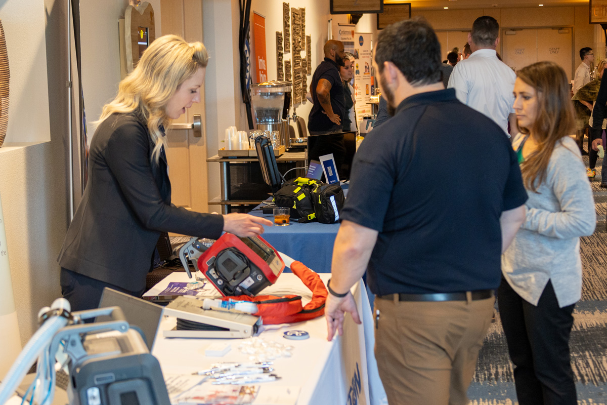
Exhibitor Information
The ALD offers dental suppliers great opportunities to sponsor or exhibit at a number of events that include single day Laser Certification courses and the Annual Session. These events have both Dentists and Hygienists who are looking for more information and training using a multitude of dental lasers and wavelengths. Click the button below for more information or call us at 813-444-1011.